Our Ophthalmologists are experts in the field of retinal disease and surgery. We take pride in delivering a wide array of comprehensive and specialized eye services to Mandeville, Metairie, and Slidell, LA.
Welcome to EyeCare 20/20 Retina & Vision Center!
Your eyes tell a unique story, and at EyeCare 20/20 Retina & Vision Center, Dr. Neil Notaroberto, MD is here to ensure that the story continues with clarity and vitality. Through our network of state-of-the-art facilities in Mandeville, Metairie, and Slidell, we’ve revolutionized the approach to vision care in Louisiana.
Our practice doesn’t just treat eye conditions – we create personalized vision solutions that enhance your daily life, whether you’re reading a beloved novel, watching your grandchild’s first steps, or pursuing your career dreams.


Our Mission
Vision is more than just seeing clearly – it’s about experiencing life’s moments in vivid detail. We’ve built our practice on the foundation of transformative eye care that goes beyond traditional boundaries.
By combining breakthrough diagnostic tools with our team’s specialized expertise, Dr. Neil Notaroberto, MD at EyeCare 20/20 Retina & Vision Center is redefining what’s possible in vision care. Each day, we witness the joy of patients rediscovering the world through improved vision, and it fuels our passion to push the boundaries of what exceptional eye care can achieve.
- Laser Cataract Surgery
- Retinal Health
- Macular Degeneration
- Diabetic Retinopathy
- Adjustable Lens
- Retinal Injection
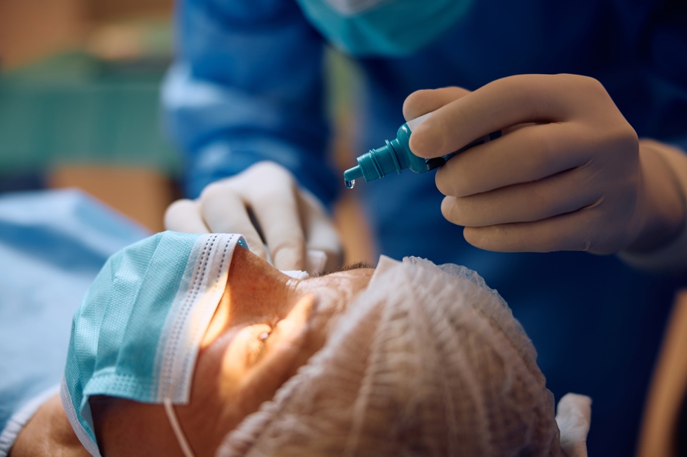
Experience Crystal Clear Vision Again with Laser Cataract Surgery
Modern eye care has transformed the way we approach cataract treatment. At EyeCare 20/20 Retina & Vision Center, our advanced laser technology delivers exceptional results for patients seeking clearer vision. The precision-guided system creates a customized treatment plan unique to your eye’s anatomy.
Understanding Your Procedure Timeline
Your journey to improved vision follows a carefully planned sequence. Our experienced team guides you through each phase, from initial consultation to post-operative care. We take time to explain every step, helping you feel confident about your decision.
- Complete eye mapping creates a detailed surgical blueprint
- Precise laser incisions take just minutes to complete
- Most patients return to daily activities within 24-48 hours
- Advanced monitoring ensures optimal healing progress
- Regular check-ups track your vision improvement
Personalized Vision Goals
Everyone’s eyes are different, and so are their vision needs. We develop specific treatment approaches based on your lifestyle and visual requirements. This individualized strategy helps achieve the best possible outcome for your unique situation.
- Custom lens selection matches your specific needs
- Advanced measurements guide precise positioning
- Digital planning enhances surgical accuracy
- Real-time adjustments optimize results
- Technology-driven approach improves consistency
Recovery and Results
The recovery process typically progresses smoothly thanks to our advanced techniques. Most patients notice vision improvements within days after surgery. Our dedicated follow-up care ensures your healing stays on track while maximizing your procedure’s benefits.
- Gentle walking permitted day after surgery
- Normal activities resume within one week
- Vision stabilizes over 3-4 weeks
- Regular check-ups monitor progress
- Detailed aftercare instructions provided
You’ll receive comprehensive instructions and support throughout your recovery. Our commitment to excellence at EyeCare 20/20 Retina & Vision Center shows in every aspect of care, from your first consultation through your final follow-up visit.
Transform Your Retinal Health Today
Specialized treatment options have revolutionized retinal care in recent years. The latest surgical techniques allow us to address complex conditions with remarkable accuracy. These procedures at EyeCare 20/20 Retina & Vision Center help protect and preserve your precious vision for years to come.
Planning Your Specialized Treatment
We begin with state-of-the-art imaging to map your retinal structure in precise detail. This thorough evaluation helps create the most effective surgical plan for your specific condition. Each step reflects our commitment to achieving optimal outcomes.
- Digital mapping creates detailed retinal images
- Custom surgical plans address specific conditions
- Advanced diagnostic tools guide treatment
- Precise measurements ensure accuracy
- Comprehensive evaluation identifies concerns
Advanced Surgical Techniques
The latest developments in microsurgical tools have enhanced our surgical capabilities. These innovations allow for more precise and less invasive procedures. EyeCare 20/20 Retina & Vision Center employs cutting-edge technology to deliver exceptional results.
- Microscopic instruments improve precision
- Latest technology minimizes tissue impact
- Advanced visualization aids surgery
- Real-time monitoring ensures safety
- Specialized tools enhance outcomes
Post-Surgery Care Excellence
Your recovery begins immediately after surgery with careful monitoring. Clear communication helps you understand what to expect during healing. We maintain close contact throughout your recovery journey.
- Detailed recovery guidelines provided
- Regular progress evaluations scheduled
- Symptom monitoring ensures safety
- Activity recommendations given
- Ongoing support available daily
A well-planned recovery program supports your healing process. Our expert team remains available to address questions and monitor your progress, helping you achieve the best possible outcome from your retinal procedure.
Advanced Solutions for Macular Degeneration
Recent medical innovations have changed how we manage macular degeneration effectively. Our comprehensive approach combines proven treatments with cutting-edge solutions. Each treatment plan focuses on maintaining your current vision while slowing condition progression.
Early Detection and Monitoring
The team at EyeCare 20/20 Retina & Vision Center uses sophisticated imaging systems to track subtle changes in your retinal health. Regular evaluations help catch early warning signs before they impact your daily activities. Quick intervention often leads to better long-term outcomes.
- High-resolution scans detect early changes
- Regular monitoring tracks progression rates
- Detailed analysis guides treatment timing
- Advanced imaging shows treatment response
- Comprehensive reports document progress
Treatment Options Available
Modern treatment methods offer new hope for managing macular degeneration. We combine different approaches based on your specific condition type and stage. Each strategy aims to preserve your remaining vision effectively.
- Injectable medications slow progression
- Light therapy targets affected areas
- Nutritional guidance supports eye health
- Preventive measures reduce risks
- Home monitoring tools track changes
Lifestyle Support Strategies
Living with macular degeneration requires certain adjustments to maintain independence. EyeCare 20/20 Retina & Vision Center provides practical solutions for everyday challenges. Our recommendations help you stay active and engaged in your favorite activities.
- Vision aids enhance daily tasks
- Lighting recommendations improve function
- Activity modifications maintain independence
- Safety strategies prevent accidents
- Environmental adjustments optimize vision
Your quality of life remains our primary focus throughout treatment. We at EyeCare 20/20 Retina & Vision Center work together to find solutions that fit your lifestyle while effectively managing your condition.
Managing Diabetic Retinopathy Successfully
Proactive diabetic eye care plays a vital role in protecting your vision long-term. Through early intervention and consistent monitoring, we identify potential issues before they affect your sight. Treatment success at EyeCare 20/20 Retina & Vision Center starts with understanding your unique health needs.
Comprehensive Disease Management
Controlling blood sugar levels directly impacts your eye health. Our specialized screening process helps track changes in your retinal blood vessels. This targeted approach allows us to develop personalized treatment strategies that work alongside your diabetes care plan.
- Blood vessel mapping guides treatment
- Regular screenings catch early changes
- Digital imaging shows vessel health
- Coordination with diabetes care
- Customized treatment timing
Latest Treatment Technologies
Modern therapeutic options provide effective ways to address retinal complications. EyeCare 20/20 Retina & Vision Center employs advanced laser systems and specialized medications. These treatments target specific areas while preserving surrounding healthy tissue.
- Precise laser treatments seal leaks
- Injectable medicines reduce swelling
- Microsurgery repairs severe damage
- Advanced imaging guides procedures
- Multiple treatment combinations available
Preventive Care Strategies
Long-term success requires an active partnership between patient and doctor. We emphasize practical steps that help maintain retinal health. Regular monitoring catches changes early when treatment works best.
- Blood sugar management tips
- Vision monitoring tools provided
- Lifestyle modifications suggested
- Regular check-up schedules
- Emergency warning signs reviewed
Your eye health remains closely connected to overall diabetes management. Our team works alongside your other healthcare providers to create a unified approach to protecting your vision.
Discover Light Adjustable Lens Benefits
Light adjustable lenses represent a breakthrough in vision correction technology. These innovative implants allow for precise vision adjustments after surgery, creating truly customized results. Our specialists at EyeCare 20/20 Retina & Vision Center use advanced light delivery devices to fine-tune your vision based on your daily activities.
Customization After Surgery
Unlike traditional lenses, LAL technology offers unique adjustment capabilities. The special light treatments modify your lens prescription while it’s already in your eye. This revolutionary approach helps achieve your specific vision goals with remarkable accuracy.
- Custom adjustments match lifestyle need
- Multiple fine-tuning sessions available
- Precise vision modifications possible
- Individual preferences considered
- Optimal results through adjustment
Light Treatment Process
Each adjustment session uses carefully controlled UV light exposure. EyeCare 20/20 Retina & Vision Center provides a series of quick, comfortable treatments. The process allows for multiple refinements until your desired vision is achieved.
- Brief treatment sessions scheduled
- Painless light delivery system
- Progressive vision improvements
- Multiple adjustment options
- Specific targeting capabilities
Post-Treatment Vision Care
Following your final adjustment, special UV protection preserves your results. Our team at EyeCare 20/20 Retina & Vision Center guides you through each phase of the process. Regular check-ups help confirm your vision stability and satisfaction.
- UV protection guidelines given
- Vision stability monitored
- Activity recommendations provided
- Progress tracking scheduled
- Long-term results maintained
The ability to customize your vision after lens implantation sets this technology apart. Your input helps shape the final results, leading to vision that truly matches your lifestyle requirements.
Understanding Retinal Injection Treatment
Modern retinal medications have created powerful options for treating eye conditions. These specialized injections deliver targeted therapy directly where it’s needed most. The expert team at EyeCare 20/20 Retina & Vision Center uses advanced techniques to maximize comfort and effectiveness during each treatment session.
Safe Treatment Administration
Professional expertise makes a significant difference in injection comfort. Our refined approach minimizes discomfort while ensuring precise medication delivery. Each session follows strict protocols to protect your eye health.
- Thorough eye preparation steps
- Precise injection techniques used
- Comfort measures implemented
- Sterile conditions maintained
- Quick procedure completion
Medication Benefits Explained
These powerful medications target specific problems within the retina. EyeCare 20/20 Retina & Vision Center selects the most appropriate option for your condition. Different medications address various retinal issues effectively.
- Specific targeting reduces swelling
- Active ingredients work quickly
- Multiple conditions addressed
- Treatment plans individualized
- Regular monitoring tracks progress
Following Your Treatment Plan
Success often depends on maintaining a consistent schedule. We create clear treatment timelines that fit your lifestyle. Regular communication helps ensure you stay on track with recommended care.
- Treatment calendars provided
- Progress closely monitored
- Side effects explained
- Activity guidelines given
- Follow-up care scheduled
Getting the most benefit from retinal injections requires good timing and proper technique. Our experienced team at EyeCare 20/20 Retina & Vision Center helps you navigate the entire process while keeping you comfortable and informed.
Our Featured Services
At EyeCare 20/20 Retina & Vision Center, we provide a comprehensive range of eye care services tailored to meet your unique needs. From routine vision care to advanced surgical treatments, our team is dedicated to protecting and enhancing your eyesight.


Advanced Technology at EyeCare 20/20 Retina & Vision Center
At EyeCare 20/20 Retina & Vision Center, we are proud to use the latest advancements in eye care technology to provide accurate diagnoses and exceptional treatment outcomes. Our state-of-the-art equipment ensures precision, safety, and effectiveness for every patient.
Procedural Equipment
Alcon’s LenSx® Laser System
Revolutionary laser technology for precise and minimally invasive cataract surgery.
Learn MoreORA System™
Enhances cataract surgery outcomes by providing real-time feedback for optimal lens placement.
Learn MoreMicro-Pulse® Laser
Advanced laser therapy for treating conditions like glaucoma with minimal tissue damage.
Learn MoreVISULAS® YAG III Laser
A trusted solution for post-cataract procedures and other laser treatments.
Learn MoreDiagnostic Imaging Equipment
- SPECTRALIS® Optical Coherence Tomography (OCT)
- Optomap® Ultra-Widefield Retinal Camera
- Retinal Angiography
- Zeiss IOL Master 700
- Diopsys VEP and ERG
- Ellex® Eye Ultrasound
- TearLab® for Dry Eye Detection
SPECTRALIS® Optical Coherence Tomography (OCT)
High-resolution imaging to detect and monitor retinal diseases.
Learn MoreOptomap® Ultra-Widefield Retinal Camera
Comprehensive imaging of the retina to detect early signs of disease.
Learn MoreRetinal Angiography
Precise imaging to evaluate blood flow in the retina and diagnose vascular issues.
Learn MoreDiopsys VEP and ERG
Non-invasive tests to assess the function of the optic nerve and retina.
Learn MoreTearLab® for Dry Eye Detection
Quick and accurate testing for diagnosing and managing dry eye syndrome.
Learn MoreWhy Choose Us
At EyeCare 20/20 Retina & Vision Center, we believe exceptional vision care requires more than just advanced equipment – it demands a deep understanding of your unique needs. Here’s what sets our practice apart.
FAQs
Do I need a referral to schedule an appointment?
In some cases, the necessity for a referral depends on the purpose of your visit and the type of insurance you carry. Please feel free to reach out to us and our team will gladly guide you through the appointment scheduling process, including any necessary prior authorizations.
When should I consider obtaining an eye exam?
While it is advisable for all healthy adults to undergo a comprehensive eye examination with an ophthalmologist at the age of 40, there are various other circumstances that may warrant a visit to our clinic:
- Suspected eye infection
- Following an acute eye injury
- Noticing changes in vision or sudden blurriness
- Experiencing new floaters or flashes in your vision
- Suffering from double vision
- Experiencing a sudden loss of vision
- Developing conditions such as diabetes, hypertension, or other systemic health issues
- Receiving a recommendation from your general health practitioner (GP) or referral from an optometrist
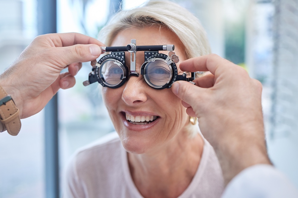
What is the difference between an eye exam and a refraction?
A comprehensive eye exam consists of a series of tests to determine overall eye health and identify potential or existing diseases of the eye that can cause vision loss. A refractive exam for glasses or contact lenses is additional testing to fit the patient in the best glasses or contact lenses for their eye and prescription.
Will my eyes be dilated during an eye exam?
Dilation is necessary for most patients to ensure a thorough exam is performed. Dilation usually affects near vision the most, leaving distance vision relatively unchanged. This means that with a good pair of sunglasses, most people are able to comfortably drive. If you are concerned about this, however, we suggest you bring someone with you to drive.
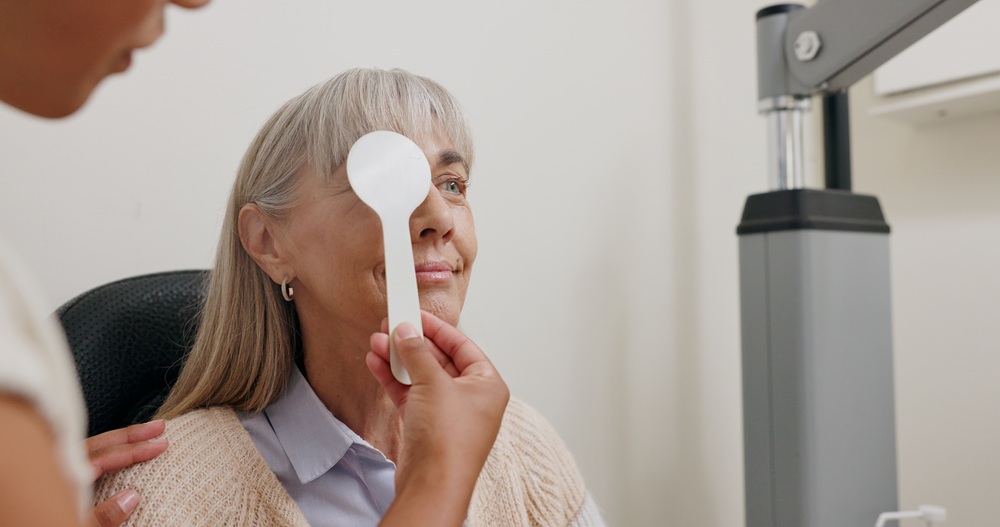
What happens during an eye exam?
During a comprehensive eye exam, your eye doctor will check your eyes for common eye diseases, assess how your eyes work together as a team and evaluate your eyes as indicators of your overall health. If requested by the patient, the eye doctor will determine if you require a prescription for eyeglasses or contact lenses.
What happens during a comprehensive eye exam?
Your exam begins with a thorough review of your medical history and vision concerns. We then perform various tests to check your visual acuity, eye pressure, and overall eye health using advanced diagnostic equipment. The appointment includes time to discuss our findings and address any questions you have about your eye health.
Do you welcome new patients at all three locations?
Yes, we actively welcome new patients at our Mandeville, Metairie, and Slidell locations. Our scheduling team will help you choose the most convenient location for your needs and guide you through any necessary paperwork before your first visit. We strive to make the process as smooth as possible for all new patients.
How do I know if my vision changes are serious?
Changes in vision like blurriness, floating spots, flashes of light, or eye pain should never be ignored. Our practice offers same-day appointments for urgent vision concerns, and our experienced team can quickly determine if your symptoms require immediate attention. We’ll provide clear guidance on managing your condition and necessary follow-up care.
What should I do to maintain healthy eyes between visits?
Protecting your eyes includes wearing UV-blocking sunglasses outdoors and taking regular breaks during screen time. We recommend following the 20-20-20 rule: every 20 minutes, look at something 20 feet away for 20 seconds. Additionally, maintaining a healthy diet and staying active can significantly benefit your eye health.
When should I seek immediate eye care?
Contact us at EyeCare 20/20 Retina & Vision Center immediately if you experience sudden vision changes, flashing lights, floating spots, eye pain, or injury. We also recommend urgent care for severe redness, light sensitivity, or if you notice a curtain-like shadow in your vision. Early intervention often leads to better outcomes for eye emergencies.
View All FAQs
Meet the Doctor
At EyeCare 20/20 Retina & Vision Center, Dr. Neil Notaroberto, MD, brings decades of specialized experience in diagnosing and treating complex eye conditions. His expertise spans from routine vision care to advanced surgical procedures, with particular focus on retinal health and cataract treatment. Dr. Notaroberto leads our practice with a simple philosophy: provide focused, individual attention to each patient while utilizing the most effective treatment options available. His dedication to staying current with medical advancements ensures that EyeCare 20/20 Retina & Vision Center patients receive the most up-to-date care for their vision needs.

Dr. Neil Notaroberto
Founder and Retinal Specialist
With additional fellowship training in retinal care, Dr. Notaroberto leads our team with a passion for innovation and patient care. His extensive experience and dedication have established our practice as one of the most respected eye care providers in Louisiana.
Dr. Arley Jaramillo
Vitreoretinal Surgeon
Known for his surgical expertise and compassionate approach, Dr. Jaramillo specializes in treating complex retinal and vision-blinding diseases. His fellowship training ensures exceptional care for even the most challenging cases.


Dr. Katie Casadaban
Board-Certified Optometrist
Dr. Casadaban provides comprehensive eye care with a strong emphasis on medical diagnoses and treatments of ocular diseases. A graduate of the Southern College of Optometry, her patient-focused approach ensures personalized care for every individual.
Dr. Anna Douglass
Board-Certified Optometrist
Dr. Douglass specializes in thorough eye exams and personalized vision solutions. With a strong foundation from LSU and the Southern College of Optometry, she is committed to helping patients achieve optimal eye health.

National Affiliations:
This Is Normal Eye Vision

Normal Vision


New Patients
Call us to schedule an appointment or find your new patient paper work here.
Existing Patients
You may access your medical records, pay/track bills, check your appointment, leave reviews, and more through your online patient portal. Learn more about Breeze or access your portal by clicking the Breeze icon below.

Testimonials
”Dr. Neil is the best in St. Tammany. I had cataract surgery & all went smooth , easy & very professional! Extra plus: he is also the nicest Dr. I ever met. He has a great office & medical staff!
Marie LMandeville
”Dr. Notaroberto is a wonderful eye doctor who takes the time needed to visit any of your concerns. Highly recommend!
Deborah MMandeville
”I would definitely recommend Dr. Notaroberto. It is amazing how my vision is so much better. His office staff are very nice.
Connie ASlidell
”Very nice staff, super clean and welcoming place! Will be back for every eye exam!
Krisa EMetairie
”The Eye Doctor here is so nice, friendly, patient, and caring. I always have such a nice experience when coming here. The other staff are very nice too.
Justin BMetairie
”Very friendly and efficient staff. Dr. Neil is always attentive and kind, explains things clearly, and goes beyond the norm to be helpful. Great office.
Don RSlidell
”Very thorough, kind, professional and has compassion for his patients, not to mention he's extremely knowledgeable in his craft. I love the staff, they are so polite and professional. I highly recommend going here for any and all retina problems.
MS JonesMetairie
”Great Doctor, explained all aspects of my condition in terms easily understood. Gave me options for treatment. I highly Recommend 20/20. Waiting time was not bad either!
Richard PSlidell

We utilize the most advanced ophthalmic diagnostic equipment available, allowing us to provide the highest quality of care.


Our team of Ophthalmologists and Optometrists are highly trained to deliver an array of eye services.
We accept all major insurances:


Count On EyeCare 20/20 Retina & Vision Center For Clear Vision!
Your vision matters to us at EyeCare 20/20 Retina & Vision Center, and we’re ready to help you maintain healthy eyes for years to come.
Connect with us on Facebook, X, and Instagram to learn about eye health tips and practice updates, or call 985-624-5058 to schedule your appointment. Whether you need a routine check-up or have concerns about your vision, Dr. Neil Notaroberto, MD will ensure you receive the attention and care you deserve.