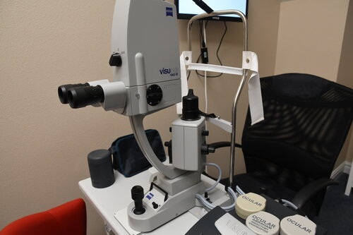
Explore our diagnostic imaging equipment


 SPECTRALIS® Optical Coherence TOmography

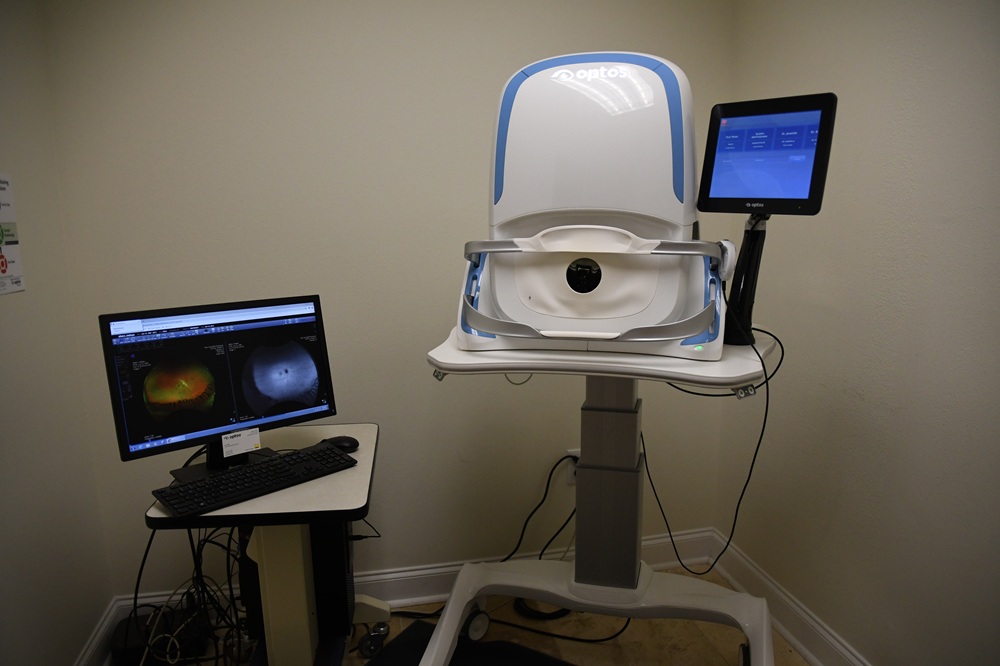
Optical coherence tomography (OCT) is a non-contact medical imaging device that uses a laser-like light source to create highly detailed cross-sectional and 3D images of the eye. This type of imaging allows us to catch early signs of eye diseases before they are detectable with traditional testing methods. Retinal changes due to macular degeneration, diabetes, and retinal vein occlusions can be detected by this form of imaging. OCT is also crucial in evaluating the optic nerve and nerve fiber layer in the management of glaucoma and other optic neuropathies.

OCT.jpg

TechThumb+-+Optos+3.jpg

Optomap® Ultra wide-field Retinal Camera

The optomap is an imaging device that takes wide-field, high resolution photos of the back of your eye. With just a quick flash, it can capture 82% of your retina in one photo, even without dilation. It is useful in documenting diseases of the retina, macula, choroid, and vitreous.


Retinal Angiography

To more closely examine the retina (the back of your eye) and choroid (blood vessels under the retina), your ophthalmologist may use a diagnostic technique called angiography. A colored dye is injected into a vein in your arm, where it travels throughout the blood vessels in your body. As the dye passes through the blood vessels in the eye, a special camera takes photographs of the retina or choroid blood vessels.Angiography helps your ophthalmologist see what is happening in your retina or choroid, highlighting any abnormalities that may be present. It is used most often to: help diagnose and monitor eye conditions including macular degeneration, diabetic retinopathy, retina vein occlusions, and macular edema; guide laser surgery with pinpoint accuracy; and monitor the effects of laser surgery. EyeCare 20/20 Retina & Vision Center is the only ophthalmology office in the area with the ability to perform extra wide field angiography.

angio.jpg

Tech - IOLMaster 5.JPG

Zeiss IOL master 700

If you suffer from cataracts, our doctors can remove your compromised lenses and replace them with artificial intraocular lenses (IOLs) during laser cataract surgery. We offer a number of IOLs to choose from, but selecting the right one can be a challenge. The Zeiss IOLMaster makes the process easier and more accurate than ever before. This state-of-the-art device quickly and accurately measures the length of your eye and the curvature of your cornea. Because of its extreme precision, our cataract surgeries offer optimal results and better vision enhancement.


Diopsys VEP and ERG

Visual Evoked Potential (VEP)

The Electroretinogram (ERG)

Diopsys.jpg

Ellex Bscan 1.JPG

Ellex® Eye Ultrasound

The Eye Cubed from Ellex is an imaging device that uses ultrasound to provide real-time imaging of the structures within the eye. It can help with the diagnosis of retinal detachments, vitreous hemorrhages, and eye tumors.


tearLab® for dry eye detection

Dry eyes are a common condition which can result from the aging process, certain medications, environmental conditions, genetics, and more. The growing use of screens and monitors can also decrease the natural blink response, which lubricates the eye. TearLab® allows our doctors to both diagnose and manage the condition. The technology is the first objective and quantitative test for dry eye syndrome, and offers fast and accurate results using samples of our patients' tears. The system allows us to pinpoint the cause of your dry eyes and offer targeted, individually tailored treatment for optimum results. 

EyeCare2020TearLab